Lines, Tubes, and Catheters in Radiography Paper
Abstract
This paper is written to explore the application and appearance of lines, tubes, catheters, as well as other medical devices in radiography. In specific, endotracheal tubes, tracheostomy tubes, drainage tubes, chest tubes, nasogastric tubes, nasoenteric tubes, and central lines or central venous lines will be included and discussed. Tubes and catheters are useful for the administration and delivery of nutrients, providing drainage routes, administering medications and fluids, drawing blood samples, and many other functions and uses. A radiographer may not be required to utilize these types of medical equipment, but to have an understanding is still important. It is necessary for patient care to have these medical devices available in order to conduct effective treatment in both trauma or non-trauma situations. Therefore, the radiographer has an imperative role in verification of the proper placement of lines, tubes, and catheters. Knowledge of the medical devices used, placement, and location within the body are essential for quality patient care.
Tubes, lines, and catheters do not always have an apparent difference. A catheter, though, iks defined as a hollow, flexible tube that can be put into a body cavity or vessel to insert or withdraw fluids (Newman, 2017). Tubes, lines, and catheters are hollow inside, and most are flexible for the removal of fluids. Other medical devices, such as cardiac pacemakers and those listed previously serve other purposes. A chest x-ray is the recommended radiograph by the American College of Radiology after tubes, lines, catheters, or other devices are inserted into the patient (Jain, 2011). It provides a go-to window to check position of equipment and other medical procedure issues. Function, location, and follow-up are what constitute the application of tubes, lines, catheters, and other devices.
An endotracheal tube is used for ventilation, maintenance of the airways, prevention of aspiration, and suction of tracheobronchial secretions (Newman, 2017). Patients who need this type of medical device are usually having trouble with their airways staying open or undergoing anesthesia. Placement for an endotracheal tube is into the mouth or nose, through the larynx, and to the trachea (Newman, 2017). An endotracheal tube consists of a terminal hole and cuff. The tube tip can be five to seven centimeters above the carina with the neck in a neutral position and two centimeters caudad or cephalad with the neck in flexion or extension, accordingly. If the carina is not visible, the tube tip should be at the medical clavicle ends and midway between the carina and larynx so extubation and intubation are evaded. A chest x-ray is necessary immediately after the tube is inserted in order to verify there is no extubation or intubation and proper positioning (Jain, 2011). Another landmark to ensure correct positioning is the aortic knob. If the endotracheal tube tip is barely above the aortic knob it is in the right spot. Incorrect placement of the tube may result in hyperinflation or collapse of the lungs (atelectasis) (Newman, 2017). Also, intubation into the esophagus indicated by an overstretched stomach and tracheal stenosis are complications to avoid (Jain, 2011). Endotracheal tubes are to be administered to the patient with precision.
A tracheostomy tube is inserted through an opening into the trachea as a result of a surgical tracheotomy for an airway (Ehrick & Coakes, 2017). This device is needed to handle an obstruction of the respiratory tract due to a cancer or burn in the mouth or throat and also for controlled breathing with a ventilator in patients who have respiratory collapse due to paralysis or trauma (Ehrick & Coakes, 2017). The placement for a tracheostomy tube is midway between the stoma and carina at the D3 vertebra and is kept with flexion and extension of the neck. Tube diameter should be two-thirds of the width of the trachea, the cuff not overstretching the tracheal wall, and the tube parallel with the trachea (Jain, 2011). Improper positioning of a tracheostomy tube can result in surgical emphysema, pneumothorax, hemorrhage, or tracheal stenosis (Jain, 2011). A chest radiography can make sure the tube is placed correctly.
A drainage tube, or pleural tube (intercostal drainage tube) is used to rid the chest of liquids and solids. It is placed through the fourth intercostal space in the anterior or mid-axillary line and then lead to go in a posteroinferior path for effusion and anterosuperior path for a pneumothorax (Jain, 2011). A chest x-ray shows the side holes on the tube in the radiopaque outline of the tube. The side holes should not be outside the chest area and the tube not hover above the effusion that may be present. Both an anteroposterior and lateral chest x-ray will verify the correct positioning of an intercostal drainage tube (Jain, 2011). Other chest tubes include thoracic catheters or thoracostomy tubes. These are used to remove air, liquid, and solids from the pleural space and mediastinum (Newman, 2017). Open suction allows for drainage into the tube due to a difference in pressure between the pleural space and atmosphere or gravity. Closed suction involves a vacuum pump and bottles. The placement for the tube is in the third to sixth intercostal space in the anterior or mid-axillary line. Again, an anteroposterior and lateral chest radiograph will indicate if the tubes are properly positioned (Newman, 2017).
Nasogastric and nasoenteric tubes are administered through the nose and down to the stomach or small intestine. Nasogastric tubes for the stomach help with feeding, decompression, and radiographic examination (Ehrich & Coakes, 2017). A nasogastric tube has many side holes and lead balls at the end of it, and its tip should be placed within the stomach about ten centimeters caudal to the gastroesophageal junction (Jain, 2011). Medication, nutrients, and contrast agents can be given to the patient with a nasogastric tube. The placement of the tube is done by a physician with informed consent from the patient. Improper positioning may result in aspiration or bowel perforation (Newman, 2017). An abdomen radiograph will visualise the location of a nasogastric tube. Nasoenteric tubes are placed in the stomach and moved into the small intestine via peristalsis (Ehrich & Coakes, 2017). A nasoenteric tube can be used to insert contrast for a radiographic evaluation. The tip of this tube is inserted about ten to twelve centimeters into the small bowe. Improper positioning can result in pneumonia, pulmonary contusion, or perforations in the pharynx or esophagus (Jain, 2011). Again, an abdomen radiograph can show if the tube is placed well.
Central lines or central venous catheters are used to administer chemotherapy or other long-term drug therapy, total parenteral nutrition, dialysis, or blood transfusions. They are also used for blood draws and checking central venous pressure (Ehrlich & Coakes, 2017). Central venous lines are used to get into the central vessels such as external jugular, internal jugular, common facial, cephalic, and saphenous vessels. A physician places a central venous catheter (Newman, 2017). The tip of the line is distal to the last venous valve where the internal jugular and subclavian veins intersect. A check x-ray will show the value inside the first rib. Improper positioning can result in pneumothorax, perforation of a vessel, or cardiac perforation (Jain, 2011). Proper placement of the central venous catheter, to monitor central venous pressure, is when the tip lies parallel to the wall of the superior vena cava, superior to the right atrium. Postprocedural central venous catheter placement requires a radiograph to confirm accuracy, because improper catheter tip location within the heart can cause cardiac perforation and tamponade (Melarkode, 2009).
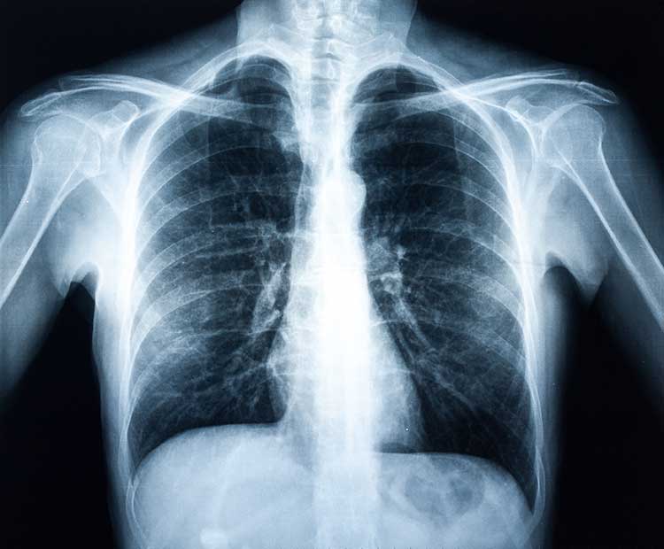
Tubes, lines, and catheters are necessary for good and effective patient care. The placement of such medical devices is particular for each one. Chest radiographs are the main indicator that correct positioning has been achieved while abdomen radiographs can also be of use. Many medical devices are not made to be seen on a radiograph (radiopaque), but some endotracheal and nasogastric tubes have radiopaque markers or tips (Newman, 2017). The design and location of a tube, line, or catheter determines the medical attention needed for the patient.
References
- Newman, J. (2017). Radiographic appearance of tubes, lines, and catheters. Radiographic appearance of patient tubes, lines, and catheters, 5(1), 1-17.
- Jain, S. (2011). A pictorial essay: Radiology of lines and tubes in the intensive care unit. Indian Journal of Radiology and Imaging, 21(3), 182.
- Erich, R. A., & Coakes, D. M. (2017). Patient care in radiography: With an introduction to medical imagine (9th ed.). St. Louis, MO: Mosby.
- Melarkode, Krishnan, and M. Y. Latoo. 2009. “Pictorial Essay: Central Venous Catheters on Chest Radiographs.” British Journal of Medical Practitioners 2 (2): 55-56
Cite This Work
To export a reference to this article please select a referencing style below:
Related Content
All TagsContent relating to: "radiography"
Radiography: specialisation in the use of radiographic, radiation therapy and magnetic resonance equipment to administer radiation treatment and produce images of body structures for the diagnosis and treatment of injury and disease.
Related Articles