Introduction
Cone beam computed tomography is the newest modality currently used in dentistry and maxillofacial radiology, delivering high quality diagnostic images. The yields images in 3-dimensional planes; axial, sagittal and coronal and is based on volumetric tomography.(1) CBCT delivers higher radiation dose than conventional dental imaging but gives dentists a clear depiction of anatomy and pathology. (2) Despite higher radiation dose, cost, poor resolution, longer scan times(3) this technology has revolutionized dentistry so much that there is hardly any dental speciality that does not require its use.(4) Incorporation of this new technology into the dental practice comes with risks and potential liabilities so clinicians should be aware of them. However, the decision to incorporate or not should not be based on legal but clinical consideration.(5)
Decision Making: Self-Referral (Own) or Referral
The decision-making process to incorporate comes with a challenging question; are the dentists ready for this technologically advanced incorporation. It is very important for dentists to consider the pros and cons of the technology. The European Academy of Maxillofacial radiology has recognized that dental undergraduate education focusses more on two-dimensional conventional dental imaging and most dental graduates receive little or no formal training in the application and interpretation of cone beam imaging.(6) The level of knowledge owing to novelty of the technique maybe insufficient to operate these sophisticated and complex machines and to meet the standards for justification, acquisition and interpretation of the images as the dentist is ultimately responsible to interpret the findings of the scan acquisition. Considering the higher dose of radiation in CBCT scans compared to conventional radiography, correct justification and interpretation is fundamental to every image scan. (7)
Financial factors should also be considered when the dentist considers incorporating this technology into their practice. CBCT machines vary in cost ranging somewhere between US $ 90,000 to in excess of US $ 300,000. This is huge investment and dentist tend to over-prescription of the scans in order to achieve return on the investment.(7) Such over prescription can leads to unethical standards in dentistry and also over exposure in the patients. Another important factor to consider is the dental space. CBCT machine come with complex mechanical components and need significant office space. Dedicated air conditioning units as well as complex subscription-based software’s are required which can lead to added cost.(8)
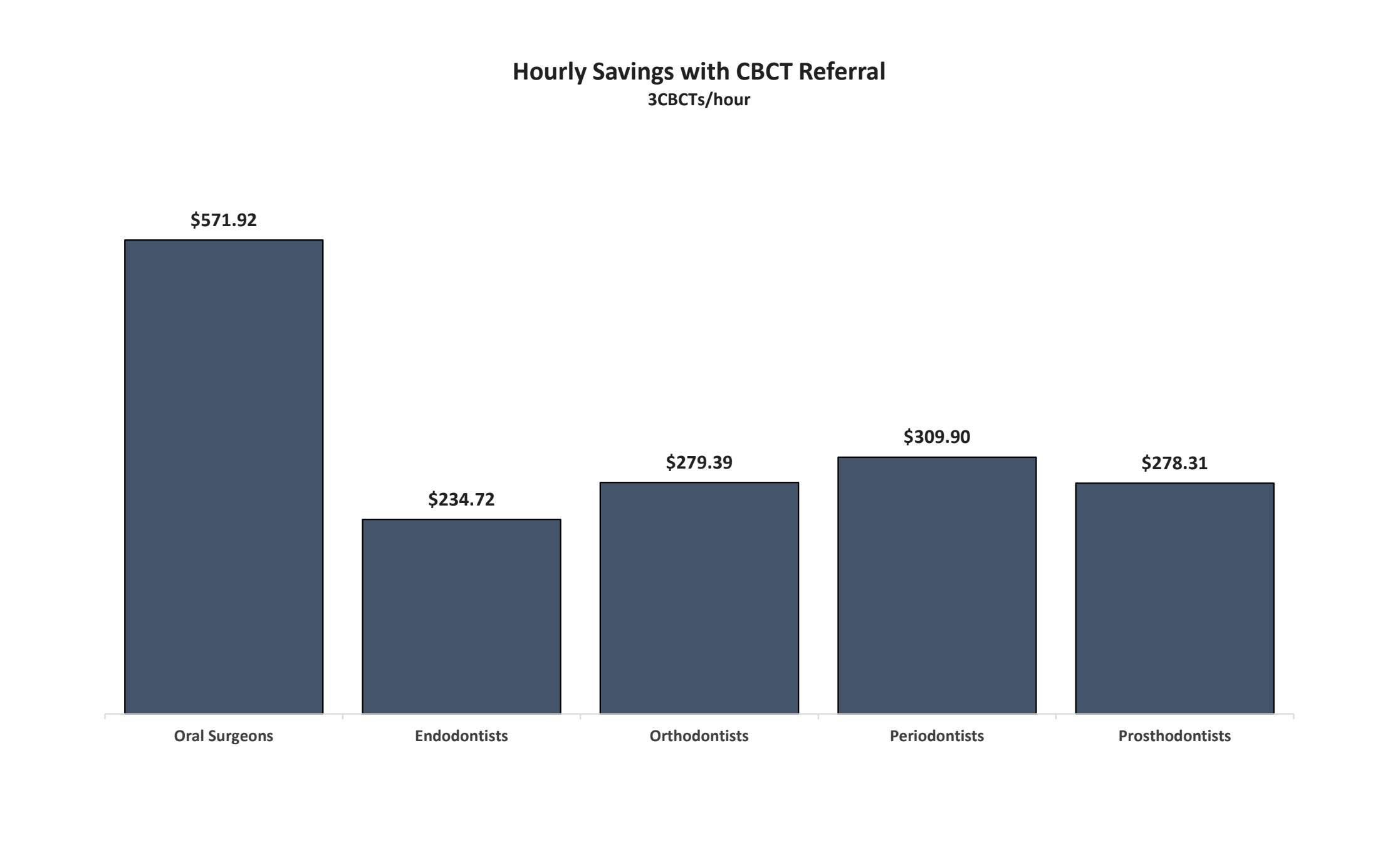
A common argument made by dentist wanting to own a CBCT machine is that they lose time and money each time they refer a scan outside to Oral and Maxillofacial Radiologist for acquisition and interpretation. However, the study has shown that it is the other way around as referral outside to Oral and Maxillofacial Radiologist is associated with significant cost savings for all dental specialities as considerable time is spent in proper interpretation of the scan which can be utilised for other important tasks(Graph 1.1).(9) Referring outside to specialist oral radiologist also come with an added advantage of providing high quality of care to their patient not to mention that the dentist who owns the CBCT is responsible for proper interpretation of the scan. The dentist is legally responsible to interpret all the anatomical area visible in the scan not just the area of interest. (5) The American Academy of Oral and Maxillofacial Radiology in its executive opinion statement states that “the “dentist using CBCT should be held to the same standards as board-certified oral and maxillofacial radiologists, just as dentists excising oral and maxillofacial lesions are held to the same standards as OMF surgeons.” (10) The referral of CBCT to outside Oral and Maxillofacial Radiologist is an efficient, high quality and cost-effective means to interpret the scans not to mention saves the dentist from legal implications.
Graph 1.1 shows the savings of CBCT referral which is determined by the difference of the hourly gross billing when referring to an Oral and Maxillofacial Radiologists vs self-interpreting CBCT scans.

What should I own as a Prosthodontics?
Osseointegration of implants has revolutionized modern prosthodontics. In addition to possessing excellent clinical skills, careful assessment of the area of implant placement and subsequent restoration is required to achieve predictable results.(4)CBCT provides significant improvement in data acquisition and a more accurate relationship of the anatomic structures as well as the bone quality and composition. Virtual planning with surgical guides predicts the final position and stability of the implants. The 3D visualization of the implant site provides more insight into the prosthetically driven implant placement, improving aesthetics and minimising the risk of treatment failures.(11) CBCT also provides clear imaging of the Temporomandibular joint area which is important consideration for full mouth rehabilitation cases which prosthodontics restore frequently.
Certain factors should be considered during the decision making process to buy a new CBCT machine such as higher image resolution, higher spatial resolution, faster scan time, low effective dose, reduced image artifacts(3), ability to select different field of view (FOV), low voxel size, beam limitation, imaging software, shorter reconstruction time, average machine cost, excellent integration with software, office space (not too bulky or heavy), Flat Panel detector type (12), good technical support and maintenance by the company.
The CBCT market has grown bigger over the past decade and a number of manufacturers are offering different models according to needs and finances. After careful research of different CBCT machine in market and based on my need as a Prosthodontists to provide quality treatment to my patient, I have chosen to buy Planmeca ProMax 3D Plus (Planmeca USA Inc.)
Planmeca ProMax 3D Plus is one of the many models of CBCT machines offered by Planmeca Inc. It is technologically the mid-level model of the various models offered and has many advantages. It is a 4-in-1 machine offering 3-D imaging along with panoramic, extraoral bitewings and cephalometric imaging. Going into the technical specifications of this machine, it has tube voltage of 54-90 KV, tube current 1-14mA which can help us to set different parameters for different patients to lower radiation dose. The scan time of this machine ranges from 9-33 secs. depending on FOV (other range somewhere between 10-40 secs) which is important for geriatric patients. Reconstruction scan time of 2-30 secs. which will decrease the wait -time for dentist before they could see the image and provide interpretation. The voxel size of 75um-400um (75um is the lowest than any other machine) provides the best resolution. FOV of this machine ranges from (4*5 to 14*9) so dentists can scan different regions based on the clinical procedure small or big and restricting the dose delivered to the patient.
The machine uses computer-controlled SCARA (Selectively Compliant Articulated Robotic Arm) which can produce accurate and reliable volume positioning and diameter adjustments reducing the amount of radiations delivered to patients. It has also intelligent 3D noise filtration which removes the noise from images without losing their quality and allows for decreased exposure values. As metal restorations and root fillings create shadows and streaks on the images, the in-built software efficiently reduces these artifacts. The machine also has Scout imaging built in to check for the patient positioning so as to prevent unnecessary high dose exposure. Other advantages of this machine are that this machine can scan both impressions and model cast which is required for Prosthetic procedures. The software integration of this machine is amazing as it can be easily integrated into Planmeca Romexis office software and implant software which can help us to plan our implant cases. Planmeca ProFace is another software that comes with the machine providing realistic 3D facial photo and CBCT image in a single imaging session which can be very useful for Prostho-Ortho interdisciplinary cases. (13)
Planmeca ProMax 3D Plus has certain disadvantages considering its high initial cost somewhere around US$ 100,000. The scan time of 9-33 secs. is still large as KaVo OP3D has scan time of 11-21secs. (14) considering the maximum scan time. The machine is pretty heavy weighing about 289 lbs., (13) however considerably low compared to other machines. So, considering the advantages and disadvantages, I would go with Planmeca ProMax 3D Plus as my choice for CBCT machine if I consider owning one for my practice. However, the decision to own depends on other important factors which we have already discussed above.
Conclusion:
CBCT provide 3D imaging of the anatomic structures and has many advantages over conventional imaging modalities. However careful considerations to made towards the legal implications, finances, training and office physical space while buying one for the dental practice. The choice of considering one machine over the other is a personal choice, but proper knowledge about technical specifications of CBCT’s machines should be done by comparing different machines and choosing the one that can provide good returns on the investments.
References
1. Hajem S, Brogårdh-Roth S, Nilsson M, Hellén-Halme K. CBCT of Swedish children and adolescents at an oral and maxillofacial radiology department. A survey of requests and indications. Acta Odontol Scand [Internet]. 2019 Aug 6 [cited 2019 Aug 28];1–7. Available from: https://www.tandfonline.com/doi/full/10.1080/00016357.2019.1645879
2. Brown J, Jacobs R, Levring Jäghagen E, Lindh C, Baksi G, Schulze D, et al. Basic training requirements for the use of dental CBCT by dentists: A position paper prepared by the European Academy of Dento Maxillo Facial Radiology. Dentomaxillofacial Radiol. 2014;
3. Venkatesh E, Venkatesh Elluru S. CONE BEAM COMPUTED TOMOGRAPHY: BASICS AND APPLICATIONS IN DENTISTRY. J Istanbul Univ Fac Dent. 2017;
4. MacDonald D. Cone-beam computed tomography and the dentist. Journal of investigative and clinical dentistry. 2017.
5. Friedland B. Liabilities and Risks of Using Cone beam Computed Tomography. Dent Clin North Am [Internet]. 2014 Jul 1 [cited 2019 Aug 28];58(3):671–85. Available from: https://www-sciencedirect-com.ezproxy.library.ubc.ca/science/article/pii/S0011853214000330
6. The 2007 Recommendations of the International Commission on Radiological Protection. ICRP publication 103. Ann ICRP. 2007;
7. Farman AG. Guest editorial--Self-referral: an ethical concern with respect to multidimensional imaging in dentistry? J Appl Oral Sci [Internet]. 2009 [cited 2019 Aug 28];17(5):i. Available from: http://www.ncbi.nlm.nih.gov/pubmed/19936508
8. Thomas SL. Application of Cone-beam CT in the Office Setting. Dent Clin North Am [Internet]. 2008 Oct 1 [cited 2019 Aug 28];52(4):753–9. Available from: https://www-sciencedirect-com.ezproxy.library.ubc.ca/science/article/pii/S0011853208000463
9. Joshua J Orgill,Suvendra Vijayan,Sindhura Anamali VA. Interactive Resource Center (IRC) The Dental Specialist’s Cost-Benefit Analysis of Referring a CBCT to an Oral and Maxillofacial Radiologist [Internet]. 2019. Available from: https://www.omfrcenter.com/manuscript1
10. Carter L, Farman AG, Geist J, Scarfe WC, Angelopoulos C, Nair MK, et al. American Academy of Oral and Maxillofacial Radiology executive opinion statement on performing and interpreting diagnostic cone beam computed tomography. Oral Surgery, Oral Med Oral Pathol Oral Radiol Endodontology [Internet]. 2008 Oct [cited 2019 Aug 28];106(4):561–2. Available from: http://www.ncbi.nlm.nih.gov/pubmed/18928899
11. Pozzi A, Arcuri L, Moy PK. The smiling scan technique: Facially driven guided surgery and prosthetics. J Prosthodont Res [Internet]. 2018 Oct 1 [cited 2019 Aug 28];62(4):514–7. Available from: https://www-sciencedirect-com.ezproxy.library.ubc.ca/science/article/pii/S188319581830015X
12. Jeff Rohde. Buyers Guide: Cone Beam 3D Imaging | Dentalcompare: Top Products. Best Practices. [Internet]. Available from: https://www.dentalcompare.com/Buyers-Guides/135269-Buyers-Guide-Cone-Beam-3D-Imaging/
13. Planmeca 3D Imaging Brochure; https://www.planmeca.com/na/imaging
14. KaVo OP 3D Brochure; https://www.kavo.com/en-us/imaging-solutions/kavo-op-3d-pro-extraoral-x-ray#docs
Cite This Work
To export a reference to this article please select a referencing style below:
Related Content
All TagsContent relating to: "dental"
Dentistry, also known as dental medicine and oral medicine, is a branch of medicine that consists of the study, diagnosis, prevention, and treatment of diseases, disorders, and conditions of the oral cavity.
Related Articles


