

General
The use of natural products with therapeutic properties is as ancient as human civilization. For a very long time, mineral, plant and animal products were the main sources of drugs [1].The use of complementary medicine to alleviate and improve health conditions is increasing in developed countries [2]. New medicinal plants from different parts of the world are being investigated with this purpose in mind [3]. Although the utilization of botanicals has increased in the western world, there is a lack of information about mechanisms of action and potential differences among species within the same genus [4].
Now-a-days, several plants have been identified for their anticancer and anti-inflammatory compounds. Scientific experiments on the anticancer properties of plants and their components have been detected. Herbal medicines have been the basis of treatment and cure for various diseases and physiological conditions in traditional methods practiced such as ayurveda, unani and siddha. However no systematic studies were conducted to evaluate the efficacy and safety of the formulations from the plant were undertaken. Also no attempts were made to isolate and identify the active principles involved in these effects [5].
As an evolutionary response plants were obliged to produce and store a wide range of organic molecules. These substances are usually termed as secondary metabolites (SM). Some of these compounds are involved in the survival of the plants as a defense mechanism against natural enemies. Many SM could actively interact with targets in the human body inducing a bioactivity of interest [6]. The bioactive compounds of medicinal plants are used as anti-diabetic, chemotherapeutic, anti-inflammatory, anti-arthritic agents where no satisfactory cure is present in modern medicines.
The use of plants for medicinal purposes dates back to antiquity because they contain components of therapeutic value [7]. Medicinal plants are cheaper and more accessible to most of the population in the world. The acceptance of traditional medicine as an alternative form of health care and the development of microbial resistance to the available antibiotics has led researchers to investigate various therapeutic uses of medicinal plants [8]. Therefore, the quest for plants with medicinal properties continues to receive attention as scientists are in need of plants, particularly of ethno botanical significance for a complete range of biological activities, which ranges from antibiotic to anti-cancerous [9].
Bangladesh features a sub-tropical climate and low-lying landmass largely adjacent to extensive river deltas. The country comprises very fertile soils and is home to some rare ecosystems such as the Sundarban’s mangrove forests. Given the fertile plains and high population density, the indigenous vegetation has mostly given way to cropland and extensive cultivation. Today, almost 60% of the landmass is used for farming, which is a global maximum value. However, originally large parts of Bangladesh featured tropical forests and marshy jungle with highly bio-diverse flora being also an excellent source for medicinal plants.
The Bangladeshi traditional medicine is a unique conglomerate of different ethnomedical influences. Due to the geographic location and sociocultural characteristics of the country, it involves traditionally rooted elements influenced by local indigenous people and close-by Indian Ayurveda and Unani medicine [10, 11]. Given its inexpensive, easily accessible and well-established health services, the use of traditional medicine is an integral part of public health services in Bangladesh with its providers being deeply embedded within the local community [12-14]. Recent data suggest that the utilization of traditional medicine health services in Bangladesh is widespread [15] and plays a crucial role in providing health care for poor people, people in rural areas and for tribal people [16].
Antioxidant and anti-inflammatory activities of medicinal plants
The anti-oxidative activity has been confirmed contributing kinds of cancer and inflammatory preventions for its multiple functional roles. The production of oxidants is a typical event associated with aerobic metabolism. When oxygen is supplied in excess or its reduction is insufficient, reactive oxygen species or free radicals such as superoxide anions, hydroxyl radicals and hydrogen peroxide are generated [17]. Accumulation of the free radicals in body organs or tissues can cause oxidative damage to biomolecules and membranes of cell, eventually leading to many chronic diseases, such as inflammatory, cancer, diabetes, aging, cardiac dysfunction and other degenerative diseases [18]. In the last 50 years, antioxidant and anti-inflammatory activities of extracts from medicinal or food plants have been extensively investigated. Many pharmacological studies have shown that extracts of some antioxidant plant possess anti-inflammatory, anti-allergic, anti-tumor, anti-bacterial, anti-mutagenic and anti-viral activities to a greater or lesser extent. Researchers reported that intake of fruits, vegetables and other foods having high antioxidant activity has been associated with reduced risks of cancer, cardiovascular disease, diabetes and other diseases [17]. Trouillas et al. investigated the antioxidant, anti-inflammatory and anti-proliferative properties of sixteen French herbal tea and found some herbs exhibited high antioxidant, anti-inflammatory and anti-proliferative activities [19]. Antioxidant activities in twenty traditional anti-inflammatory herbs extracts were investigated. The results suggested that the anti-inflammatory activities of these extracts could be explained, at least in part, by their antioxidant properties [20]. Free radicals liberated from phagocyte cells are important in inflammatory processes, because they are implicated in the activation of nuclear factor kB, which induces the transcription of inflammatory cytokines and cyclooxygenase-2 [21].
Free radicals and oxidative stress
Reactive oxygen species (ROS) is a term that encompasses all highly reactive, oxygen containing molecules, including free radicals. Types of ROS include the hydroxyl radical, the superoxide anion radical, hydrogen peroxide, singlet oxygen, nitric oxide radical, hypochlorite radical, and various lipid peroxides. All are capable of reacting with membrane lipids, nucleic acids, proteins and enzymes, and other small molecules, resulting in cellular damage. ROS are generated by a number of pathways. Most of the oxidants produced by cells occur as:
- A consequence of normal aerobic metabolism: approximately 90% of the oxygen utilized by the cell is consumed by the mitochondrial electron transport system.
- Oxidative burst from phagocytes (white blood cells) as part of the mechanism by which bacteria and viruses are killed, and by which foreign proteins (antigens) are denatured.
- Xenobiotic metabolism, i.e., detoxification of toxic substances.
Consequently, things like vigorous exercise, which accelerates cellular metabolism; chronic inflammation, infections, and other illnesses; exposure to allergens and the presence of “leaky gut” syndrome; and exposure to drugs or toxins such as cigarette smoke, pollution, pesticides, and insecticides may all contribute to an increase in the body’s oxidant load [22].
Most reactive oxygen species are generated as by-products during mitochondrial electron transport. In addition ROS are formed as necessary intermediates of metal catalyzed oxidation reactions. Atomic oxygen has two unpaired electrons in separate orbits in its outer electron shell. This electron structure makes oxygen susceptible to radical formation. The sequential reduction of oxygen through the addition of electrons leads to the formation of a number of ROS including: superoxide; hydrogen peroxide; hydroxyl radical; hydroxyl ion; and nitric oxide.
Free radicals and other ROS are derived either from normal essential metabolic processes in the human body or from external sources such as exposure to X-rays, ozone, cigarette smoking, air pollutants, and industrial chemicals. Free radical formation occurs continuously in the cells as a consequence of both enzymatic and non-enzymatic reactions. Enzymatic reactions, which serve as source of free radicals, include those involved in the respiratory chain, in phagocytosis, in prostaglandin synthesis, and in the cytochrome P-450 system. Free radicals can also be formed in non-enzymatic reactions of oxygen with organic compounds as well as those initiated by ionizing reactions.
Some internally generated sources of free radicals are – Mitochondria, Xanthine oxidase, Peroxisomes, Inflammation, Phagocytosis, Arachidonate pathways, Exercise, Ischemia/reperfusion injury etc.
Some externally generated sources of free radicals are- Cigarette smoke, Environmental pollutants, Radiation, Certain drugs, pesticides, Industrial solvents, Ozone etc.
Normally, cells defend themselves against ROS damage with enzymes such as alpha-1-microglobulin, superoxide dismutases, catalases, lactoperoxidases, glutathione peroxidases and peroxiredoxins. Small molecule antioxidants such as ascorbic acid (vitamin C), tocopherol (vitamin E), uric acid, and glutathione also play important roles as cellular antioxidants. In a similar manner, polyphenol antioxidants assist in preventing ROS damage by scavenging free radicals. In contrast, the antioxidant ability of the extracellular space is less – e.g., the most important plasma antioxidant in humans is uric acid.
Effects of ROS on cell metabolism are well documented in a variety of species. These include not only roles in apoptosis (programmed cell death) but also positive effects such as the induction of host defense genes and mobilization of ion transport systems. This implicates them in control of cellular function. In particular, platelets involved in wound repair and blood homeostasis release ROS to recruit additional platelets to sites of injury. These also provide a link to the adaptive immune system via the recruitment of leukocytes.
Reactive oxygen species are implicated in cellular activity to a variety of inflammatory responses including cardiovascular disease. They may also be involved in hearing impairment via cochlear damage induced by elevated sound levels, in ototoxicity of drugs such as cisplatin, and in congenital deafness in both animals and humans. ROS are also implicated in mediation of apoptosis or programmed cell death and ischemic injury. Specific examples include stroke and heart attack.
All the biological molecules present in our body are at risk of being attacked by free radicals. Such damaged molecules can impair cell functions and even lead to cell death eventually resulting in diseased states.
In recent years it has become apparent that the oxidation of lipids, or lipid peroxidation, is a crucial step in the pathogenesis of several disease states in adult and infant patients. Lipid peroxidation is a process generated naturally in small amounts in the body, mainly by the effect of several reactive oxygen species (hydroxyl radical, hydrogen peroxide etc.). It can also be generated by the action of several phagocytes. These reactive oxygen species readily attack the polyunsaturated fatty acids of the fatty acid membrane, initiating a self-propagating chain reaction. The destruction of membrane lipids and the end-products of such lipid peroxidation reactions are especially dangerous for the viability of cells, even tissues [23-25].
Membrane lipids present in subcellular organelles are highly susceptible to free radical damage. Lipids when reacted with free radicals can undergo the highly damaging chain reaction of lipid peroxidation (LP) leading to both direct and indirect effects. During LP a large number of toxic byproducts are also formed that can have effects at a site away from the area of generation, behaving as ‘second messengers’. The damage caused by LP is highly detrimental to the functioning of the cell [26].
Lipid peroxidation is a free radical mediated process. Initiation of a peroxidative sequence is due to the attack by any species, which can abstract a hydrogen atom from a methylene group (CH2), leaving behind an unpaired electron on the carbon atom (•CH). The resultant carbon radical is stabilized by molecular rearrangement to produce a conjugated diene, which then can react with an oxygen molecule to give a lipid peroxyl radical (LOO•). These radicals can further abstract hydrogen atoms from other lipid molecules to form lipid hydroperoxides (LOOH) and at the same time propagate LP further.
The process of LP, gives rise to many products of toxicological interest like malondialdehyde (MDA), 4-hydroxynonenal (4-HNE) and various 2-alkenals. Isoprostanes are unique products of lipid peroxidation of arachidonic acid and recently tests such as mass spectrometry and ELISA-assay kits are available to detect isoprostanes [27].
Oxidation of proteins by ROS/RNS can generate a range of stable as well as reactive products such as protein hydroperoxides that can generate additional radicals particularly upon interaction with transition metal ions. Although most oxidized proteins that are functionally inactive are rapidly removed, some can gradually accumulate with time and thereby contribute to the damage associated with ageing as well as various diseases. Lipofuscin, an aggregate of peroxidized lipids and proteins accumulates in lysosomes of aged cells and brain cells of patients with Alzheimer’s disease [28].
Inflammation
Inflammation is one of the body unique mechanisms that help body to protect itself against infection, burn, toxic chemicals, allergens or other noxious stimuli [29]. It is a body defense reaction in order to eliminate or limit the spread of injurious agent [30]. The process is created by immune cells invading the tissue like an army in full battle mode [31].
There are various components of inflammatory reaction that can contribute to the associated symptoms and tissue injury [30]. During inflammation, innate cells and molecules are usually stimulated to isolate, destroy infectious agents and repair tissue, or sometimes the adaptive immune system is also stimulated [32]. Consequently, the mechanism works in a cascade, where the inflammation is often triggered by circulating immune complexes that enter tissues [31].
Principally, inflammation is manifested by pain, swelling, redness band loss of function in the afflicted tissue [31, 33]. Saladin (2007) categorized process of inflammation into three major processes; mobilization of the body’s defenses, containment and destruction of pathogens, and tissue clean up and repair [34]. While Mahat and Patil (2007) classified the process into three phases; the first phase is caused by an increase in vascular permeability, the second one by infiltration of leukocytes and the third one by granuloma formation [30].
The inflammatory response is initiated by circulating proteins and blood cells when they contact invaders in the tissue. Microbial invaders that lodge in body tissue and begin to proliferated triggered inflammatory response [33]. Bacterial products interact with plasma factors and cells to produce agents that attract neutrophils to the infected area (chemotaxis). The chemotactic agents, which are part of a large family of chemokines, include a component of the complement system (C5a), histamine, kinins, leukotrienes, and polypeptides from lymphocytes, mast cells, and basophils [35]. The neutrophils also produce oxidants and release granular constituents comprising of lytic enzymes performing important role in inflammatory injury [36]. The innate immune system contributes to inflammation by activating the alternative and lechitin-binding complement pathways, attracting and activating phagocytic cells that secrete cytokines and chemokines, activating NK cells, altering vas. The result would firstly be increased in blood flow to the affected tissue which accelerates the delivery of immune system element to the site [33]. The vasodilation would later cause enlarged capillaries and lead to redness (erythema) as well as increase in temperature, which for an influx of fluid and cells, contributing to swelling [32]. Saladin (2007) explained that the increased in blood flow also washes toxin and metabolic wastes from the tissue rapidly. In addition, vasoactive chemicals cause endothelial cells of the blood capillaries to separate a little, widening the intracellular cleft between them and increasing capillary permeability that ease the movement of fluid, leukocytes, and plasma proteins from the bloodstream into the surrounding tissue [34].
In the area of injury, many of the neutrophils enter the tissues. As neutrophils encounter bacteria, they avidly phagocytize, digest and destroy them. Neutrophils also recruit macrophages and additional neutrophils by secreting cytokines [33]. Activated macrophages and T cells in the inflamed tissue also secrete cytokines called colony stimulation factors, which promotes the production of more leukocytes by the red bone marrow. Within a few hours of inflammation, neutrophilia (the rise in the neutrophil count in blood) would occur [37].
Then, the neutrophils are attracted to the endothelial surface by selectins, and they roll along it. They bind firmly to neutrophil adhesion molecules of the integrin family. They next insinuate themselves through the walls of the capillaries between endothelial cells by a process called diapedesis [35]. Leukocytes adhere loosely to the selectins and slowly tumble along the endothelium, sometimes coating it so thick that they obstruct blood flow. This adherence to the vessel wall is called margination [38].
Later, the fibrinogens are filtered into the tissue fluid clots in area adjacent to the injury, forming a sticky mesh that sequesters bacteria and other microbes [34]. This is caused by release of chemicals from tissues and migrating cells. Most strongly implicated are the prostaglandins (PGs), leukotrienes (LTs), histamine, bradykinin, platelet- activating factor (PAF) and interleukin-1 [39]. Prostaglandin is implicated in inducing the production of various chemo-attractants and pro-inflammatory cytokines [36]. Gislason (2009) mentioned that macrophages and neutrophils are responsible to secrete a number of mediators which is responsible not just for initiation, but also for progression and persistence of acute or chronic state of inflammation [31].
Finally, monocytes acts as the major agent in tissue clean up and repair. It enters the blood from the bone marrow and circulated for about 72 hours. Then, they enter the tissues and become tissue macrophage. The macrophage becomes activated by lympokines from T lymphocytes [35]. The activated macrophage migrate in respond to chemotactic stimuli and later engulf and destroy bacteria, damaged host cells, as well as dead and dying neutrophils. Besides that, it also acts as antigen presenting cells and activating specific immune response [34].
Edema may also contribute to the tissue clean up. Nitric oxide is responsible for vasodilatation, increase in vascular permeability and edema formation at the site of inflammation [36]. The swelling compresses veins and reduce venous drainage, while it forces open the valve of lympathic capillaries and promote lympathic drainage. The lymphatics can collect and remove bacteria, dead cells, proteins and tissue debris better than blood capillary can. An accumulation of dead cells of neutrophils with other debris tissue and fluid will form pus, a yellowish fluid. It may accumulate in the tissue cavity and known as abcess [32]. Pus is usually absorbed, but sometimes it may be released by its rupture. Blood platelets and the endothelial cells in an area of injury secrete platelet derived growth factor, an agent that stimulates fibroblast to multiply and synthesize collagen. Hyperemia at the same time delivers the oxygen, amino acids and other necessities of protein synthesis, while the heat of inflamed tissue increases metabolic rate and speed of mitosis and tissue repair. The fibrin clot in inflamed tissues may provide a scaffold for tissue reconstruction [34].
In part, inflammation declines simply because the mediators of inflammation have short half-lives, are degraded after their release, and are produced in quick bursts, only as long as the stimulus persists. In addition as inflammation develops, the process also triggers a variety of stop signals that serve to actively terminate the reaction [38].
Anti-inflammatory Activity
Anti-inflammatory refers to the property of a substance or treatment that reduces inflammation. Anti-inflammatory drugs make up about half of analgesics, remedying pain by reducing inflammation as opposed to opioids, which affect the central nervous system.
Drugs to control inflammation
When healing is complete, the inflammatory process usually subsides [32]. However, an uncontrolled and persistent inflammation that sometimes is triggered by harmless agent such pollen or by an auto immune response. It may act as an etiologic factor for many of these chronic illnesses, where it may induce, maintain or aggravate the disease [29]. As mentioned, the inflammation would occur with the presence of antigen.
Thus, constant supply of antigen is available from the food or environment may leads to chronic inflammation and causes diseases such as asthma, arthritis and other autoimmune diseases [31]. In such cases, the defense reaction themselves may cause progressive tissue injury. Hence, anti-inflammatory or immunosuppressive drugs may be necessary to modulate the inflammatory process [37].
Anti-inflammatory drugs are designed to targets the inhibition of the release of these mediators to control inflammation [36]. Harvey and Champe (2008) have classified anti-inflammatory drugs into three category; Nonsteroidal anti-inflammatory drugs (NSAIDs), Cyclooxygenase-2 inhibitors (COX-2 inhibitors) and other analgesics [32].
Aspirin is a prototype of traditional NSAIDs. It works by irreversibly inhibit Cyclooxygenase 1 and 2 (COX-1 and 2) enzymes, which results in decreased formation of prostaglandin precursors [40]. Due to this mechanism of action, aspirin also cause adverse effects such as gastric hemorrhages, hypersensitivity and thrombocytopenia [41]. It is becoming a concern of healthcare providers that patients are developing intolerance from day to day. About fifteen percent of patients show intolerance with aspirin. Therefore, newer NSAIDs with greater anti-inflammatory activities are developed. However, the newer NSAIDs are considerably more expensive than aspirin and some have proved to be more toxic in the other way [32].
The second category is COX-2 inhibitor. The mechanism of action is by selectively inhibiting the activity of COX-2 enzyme that results in decreased of prostaglandin precursors [37]. Unlike aspirin, COX-2 inhibitors have an advantage by showing lower risk of developing gastrointestinal bleeding and have no significant effects on platelets [42]. However, this drug is not recommended for renal impaired patients because it may cause renal insufficiency and increase the risk of hypertension [40]. It also has some possible adverse effects recorded in Malaysian Index of Medical Specialities (MIMS) that it may cause allergic reaction, dizziness, headache, rash, upper respiratory infection and gastrointestinal disturbances such as dyspepsia, abdominal pain and diarrheas [41].
Acetaminophen is categorized under other analgesic because it has little or no anti-inflammatory activity [32]. It inhibits the synthesis of prostaglandin in the central nervous system and peripherally blocks pain impulse generations [40]. They have therapeutic advantages over narcotic analgesics which they do not cause physical dependence or tolerance and does not affect platelet function or increase blood clotting time, but it does have many of side effects similar to aspirin [42]. Rarely, skin rash and allergic reaction may appear as the side effects [41].
There are also drugs from autacoids antagonist such as antihistamines used to prevent progress of inflammation. The term antihistamine, without a modifying adjective, refers to the classic H1 receptor blockers [32]. H1 histamine antagonists drugs are develop effectively to target the receptors to treat hay fever and some skin allergies such as urticaria. H1 receptor blockers act on immunoglobulin E (IgE) antibody-sensitizing mast cell [37].
Rheumatoid arthritis is alleviated by drugs, which inhibit the cyclooxygenase enzyme and reduce synthesis of prostanoids, corticosteroids prevent the formation of both prostaglandins and leukotriens by causing the release of lipocortin that leads to inhibition of phospholipase A2 that reduces arachidonic acid release which is able to suppress the inflammation of rheumatoid arthritis and asthma [39]. Anti-cytokines therapy involving target on Interleukin-1b (IL-1b) and tumor necrosis factor-α (TNF-α) that stimulate synovial cells to proliferate and synthesize collagenase, leading to degradation of cartilage, stimulation of bone resorption, and inhibition of proteoglycan synthesis is another method that is effective in treating rheumatoid arthritis [32].
Despite the benefits that the drugs hold, it also carries the side effects. As a result, it may lead various unwanted effects such as to gastric lesions, allergy reactions, tolerance and dependence, as well as resistance [40, 43]. Hence, worldwide researchers are still working to produce the ideal medicines of anti-inflammatory with highest efficacy, best potency and lowest or none side effects.
- Description of the plant investigated
Bauhinia acuminata L. is a species of flowering shrub native to tropical southeastern Asia. The exact native range is obscure due to extensive cultivation, but probably from Malaysia, Indonesia (Java, Borneo, Kalimantan, Lesser Sunda Islands), and the Philippines.
It is widely cultivated throughout the tropics as an ornamental plant. It may be found as an escape from cultivation in some areas, and has become naturalized on the Cape York Peninsula, Australia [44].
Common Name
Bangla: Shwet Kanchan.
Malaysian: Bunga Perak.
English: Dwarf White Bauhinia, White Mountain Ebony [45].
Indonesian: Panawar Saribu (Sunda Islands); Kupu-kupu (Java)
Thailand: Ka Long, SomSio
Burma: Mahahlegabyu
India: Kaanchnaara, Kovidaara (Ayurvedic); Kachnaal (Unani); Vellaimandarai (Siddha/Tamil); Kanchan (Assam); Shwetkachnar, Kachnalsafaid (Punjab)
Sri Lanka: Sudu Kobalila (Singhalese)
Japan: Moku-wan-ju.
Taxonomical Hierchy:
|
Kingdom |
: Plantae |
|
Subkingdom |
: Viridaeplantae |
|
Infrakingdom |
: Streptophyta |
|
Division |
: Tracheophyta |
|
Subdivision |
: Spermatophytina |
|
Infradivision |
: Angiosperms |
|
Class |
: Eudicots |
|
Superorder |
: Rosids |
|
Order |
: Fabales |
|
Family |
: Fabaceae |
|
Subfamily |
: Caesalpiniaceae |
|
Genus |
: Bauhinia |
|
Species |
: B. acuminata |
|
Binomial name |
: Bauhinia acuminata L. |
Botanical Description:
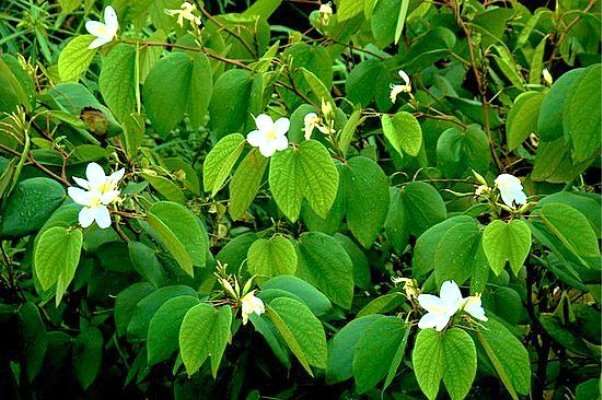
Fig.-1.1: Bauhinia acuminata L. tree.
Bauhinia acuminata L. is a member of the Fabaceae family. It is a rapidly growing shrub that can reach up to 3m tall. It rises with several strong, smooth, upright stems with many slender branches; young twigs being pubescent. The stipules are linear-lanceolate measures 1cm long. The leaves are cordate or nearly so are the base, bilobed to about one third of their length with obtuse or acute lobes 9-11 nerve, sparsely pubescent beneath, about 10cm long and broad. The flowers appear at the extremities of the branches 3-4 in a loose bunch with white petals. Thepedicels measure 6-12mm long. The flower buds fusiform, long attenuate at the apex and 5 setaceous dents, measures 3cm long. The calyx-limb laterally splitting, spathaceous; receptacle short. The petals obovate, measure 4cm long and 2cm wide. The stamens 10 all fertile, shorter than the petals; anthers small.The ovary shortly stipitate, sparsely pubescent. The pods are linear-oblong, stipitate, measure 10cm long and 1.5cm wide, dark brown in colour containing 10 roundish compressed seeds [46, 47].
Traditional uses of Bauhinia acuminata L.
The bark and leaves in a decoction helps relieve biliousness [48]. A remedy recommended by the Indian Vaiydas [45]. In Malaysia and Indonesia the plant is used in the treatment of common cold and cough [48]. The root seems to be the part made used of by the Japanese in treating cough and cold. In India the decoction of the leaves and bark is given for allying asthmatic attack. The Indians made used of the bark and leaves in a decoction to treat stones in the bladder, venereal diseases and leprosy [45]. Amongst the Mullu kuruma tribe of Karella the decoction of the bark is used in treating urinary discharge (gonorrhea). They make use of paste of the leaves applied on the throat for throat troubles. It is applied externally to treat skin diseases [49]. The root is boiled in oil and applied to burns and pain [45].
Objective of study
To evaluate the free radical scavenging and anti-inflammatory activity of Bauhinia acuminata L. bark extracts in rats.
Significance of study
Bauhinia acuminata L. is one of the
Cite This Work
To export a reference to this article please select a referencing style below:
Related Content
All TagsContent relating to: "rheumatoid arthritis"
Rheumatoid arthritis (RA) is an autoimmune disease of the joints (i.e., inflammatory arthritis [IA]) that inflames the body. RA affects 1-2% of the world’s population and is two to three times greater in women than men. RA can present itself at any age, yet statistics have shown it is more common during an individual’s third to sixth decades
Related Articles
