Paget’s Disease Description, Epdemology and Treatment
Abstract
In an average healthy adult, bones within the body are constantly regenerating and strengthening certain areas. When it comes to those with Paget’s disease, also known as Osteitis Deformans, that is not the case. It is a rare condition in which cells in the body regenerate faster than necessary resulting in a product of poor quality. There cause of the condition is still yet to be determined and there is no cure. With Paget’s disease comes pain that can be managed and the life expectancy after diagnosis is high (Altman, Udell, 2017).
Paget’s Disease
Description of Pathology
Paget’s disease is a condition within the skeletal system that effects how bones rebuild and the rate at which this occurs. Usually, the older you get the slower it will occur, but it is not the case here. This process can cause the bones to become excessively dense in some areas and weak in others due to such a rapid restoration. For the most part this disease only arises in those that are over the age of 40, but the symptoms are often overlooked due to the fact that they are expected to be linked with a certain age group (Altman, Udell, 2017).
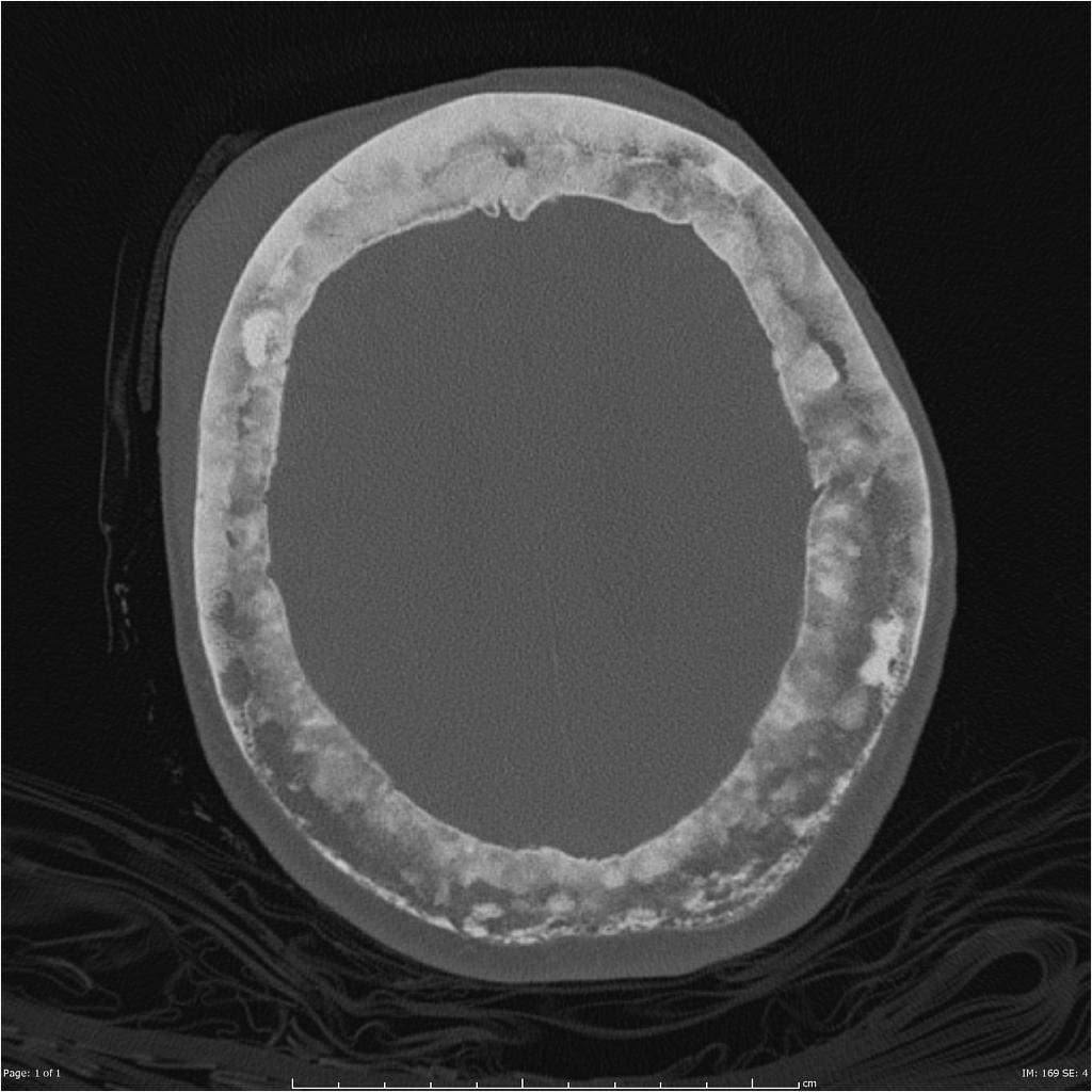
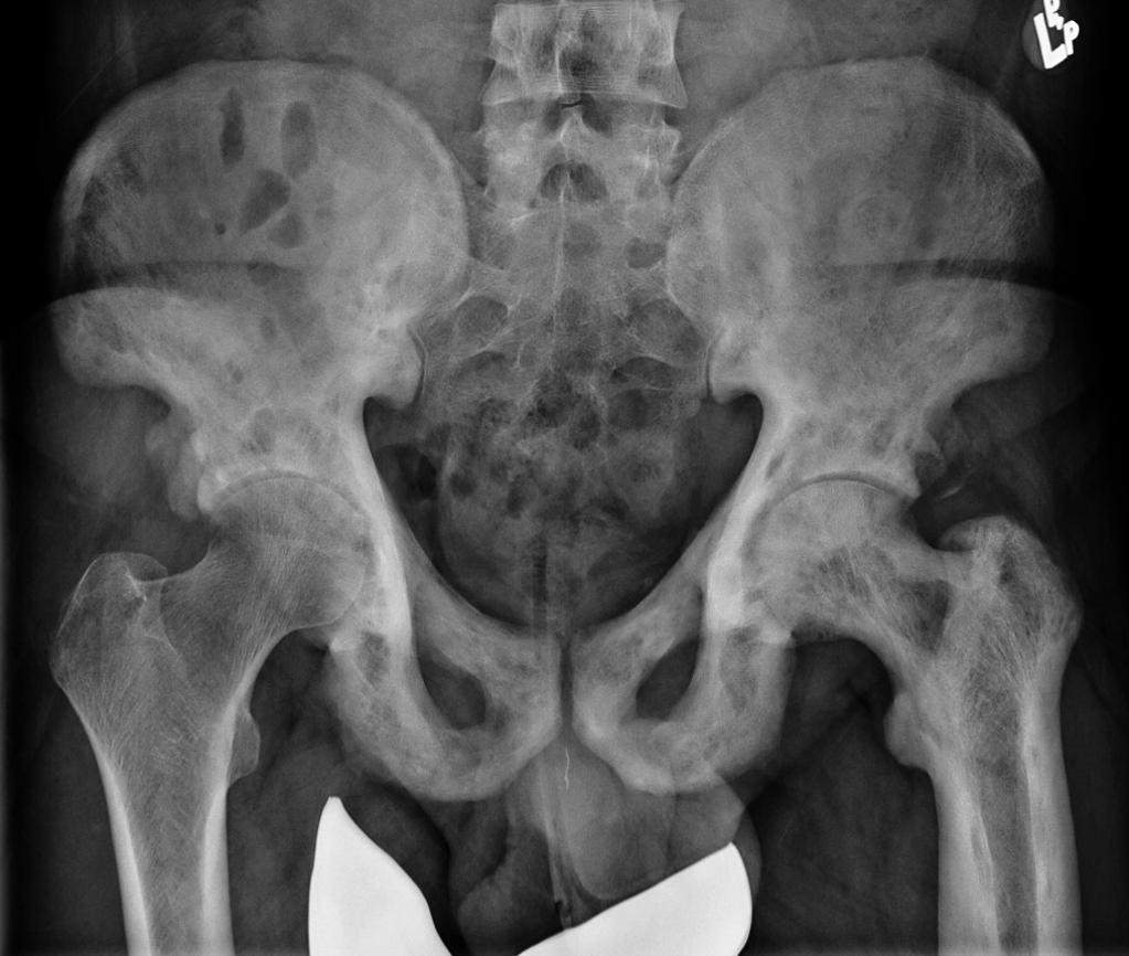
Anatomy. Osteitis deformans can be found in the spine, skull, pelvis and long bones of limbs. It may only be present in one location and only a section of it, or it can be found numerous locations. In the skull of figure 1 of the skull two rings are present. This area of interest normally would not look like this because the rings would be closer together. The space between, also known as the cortex has areas that are sclerotic, meaning thick or rigid and lucent, meaning less dense. This is a characteristic that is present in this disease it is often referred to as “cotton wool spots” (Gillard, 2019). In the pelvis of figure 2 it is present that there is an inconsistency between the sclerotic and lucent area of the hips and proximal femurs. There is a distinct medical abnormality in terms of density occurring near the acetabulum bilaterally and along the shaft of the left femur (Skalski, 2019).
Characteristics. Normally, bones to go through a process of remodeling to make certain areas stronger when they endure a repeated stress. Osteoclasts are cells that absorb bone and osteoblasts are cells that create new bone. The problem that occurs with this condition is that the osteoclasts are too active. As a result, osteoblasts aren’t working at the same pace and overcompensate by over producing bone that is abnormal in shape and size. It has been compared to a mosaic pattern in terms of structural integrity. The idea behind it is even though the pieces fit together, and the bond looks strong, but it is actually weak in how it was put together (Rajani, Fischer, 2018).
Epidemiology.
Prevalence. As of 2017, Paget’s disease is estimated to affects about one percent of the population within the United States of America (Altman, Udell, 2017).
Incidence. A study done in 2002 in England and Wales looked into the incidence of the condition to assess the correlation of age and gender in adults. The study found that men were primarily afflicted more often than woman over the age of 55 (Nebot Valenzuela, Pietschmann, 2016).
Frequency. It has been found that Paget’s occurs less frequently in women than in men, with a ration of 2:3. Correlations have been made that it occurs more often in European populations and future generations. The chance of several family members presenting with signs and symptoms of the disease was shown in 30% of cases (Altman, Udell, 2017).
Classification. Osteitis deformans is considered a chronic bone disease. The signs and symptoms are rarely seen before the age of 40, but they can occur (Altman, Udell, 2017).
Causation. While correlations have been made to determine if it is hereditary, it has not been proven yet. Currently, research is being done to look into the gene that scientist believe it related to Paget’s. If the gene can be identified they can determine who is at risk and if there are any preventable measures that can be taken (Altman, Udell, 2017).
Diagnosis
Symptoms. Due to the late onset of this condition symptoms are not present. Once they begin one can expect bone pain at or around the joints, advanced arthritis, loss of sensation in extremities and loss of movement of extremities. In extremely rare cases due to elevated calcium levels in the blood a patient can expect fatigue, weakness, decrease appetite and constipation (Rajani, Fischer, 2018).
Signs. Once the disease is in full swing joint spaces will narrow or become nonexistent. Fractures will occur due to brittleness of the bone. Bone deformities such as bowing can also be present (Rajani, Fischer, 2018). Hearing loss can occur as well, which is not reversible (Altman, Udell, 2017).
Manifestations. One percent of patients experience an extremely rare manifestation in which the disease progresses into a bone cancer called Paget’s sarcoma. If this occurs the patient will feel intense continuous pain to the area effected by the disease. This generally happens to patients over the age of 70, the tumor is aggressive, and the prognosis is poor. At this time there is no way to lessen the risk of developing the sarcoma (Rajani, Fischer, 2018).
Tests. The tests to diagnosis this condition would be x-rays, blood test and urine tests. X-rays are used to check the density of the bone, but in the early stage it may appear as hole in the bone rather than light and dark areas. The blood and urine tests look into serum alkaline phosphatase levels being increased. This shows if there is a high turnover rate in bone production (Rajani, Fischer, 2018).
Procedures. Procedures that can be useful are bone scans and biopsies. Bone scans are used to identify which areas are being affected. This is done by inserting radioactive contrast into the body to look for “hot spots” which is increased bone production. If the patient has had the condition for a long time, this test is not necessarily helpful because they can be “burned out”. Most likely meaning that the condition is too widespread. Biopsies are used to confirm the condition (Rajani, Fischer, 2018).
Treatment
Treatment for this condition is limited in the aspect of treat the symptoms, not the condition. Medication is prescribed for individuals to combat pain and swelling. This can be oral or injectable medications. Surgery is an option to combat the arthritis, which in turn can improve mobility. Assistive devices such as shoe wedges, canes, walkers and physical therapy can be provided for additional assistance (Altman, Udell, 2017).
Prognosis. The outlook for Paget’s disease is good when symptoms are managed. The patient can live a relatively normal life. As for Paget’s sarcoma does not have a good outlook and can affect the patient’s overall health (Shiel, Driver, 2018).
References
- Altman, R., & Udell, J. (2017, March). Paget’s Disease of Bone. Retrieved from https://www.rheumatology.org/I-Am-A/Patient-Caregiver/Diseases-Conditions/Pagets-Disease-of-Bone
- Gillard, F. (2019). Paget Disease of the Skull 1 [Digital image]. Retrieved from https://radiopaedia.org/articles/paget-disease-bone?lang=us
- Nebot Valenzuela, E., & Pietschmann, P. (2016). Epidemiology and pathology of Paget’s disease of bone – a reviewEpidemiologie und Pathologie des Morbus Paget – ein Überblick. Wiener medizinische Wochenschrift (1946), 167(1-2), 2-8.
- Rajani, R., & Fischer, S. J. (2018, April). Our knowledge of orthopaedics. Your best health. Retrieved from https://orthoinfo.aaos.org/en/diseases–conditions/pagets-disease-of-bone
- Shiel, W. C., Jr., & Driver, C. B. (18, May 3). Paget’s Disease Treatment, Symptoms & Diagnosis. Retrieved from https://www.medicinenet.com/pagets_disease/article.htm#paget#39s_disease_facts
- Skalski, M. (2019). Polyostotic Paget Disease 1 [Digital image]. Retrieved from https://radiopaedia.org/articles/paget-disease-bone?lang=us

Figure 1. Case courtesy of A.Prof Frank Gaillard, Radiopaedia.org, rID: 2639

Figure 2. Case courtesy of Dr Matt Skalski, Radiopaedia.org, rID: 25126
Cite This Work
To export a reference to this article please select a referencing style below:

